Ultrasound is often the first scan a GP will arrange when something feels off. Our mobile team runs scans across Brisbane, Ipswich, Logan, the Redlands and parts of Moreton Bay, so we wanted to lay out what the scan can tell you about lumps and masses, and where it stops being useful.
Short Answer
Yes, ultrasound finds plenty of tumours, although how well it picks them up depends on where the mass sits, how big it is, and what it's made of. For a firm diagnosis you'll usually need a biopsy as well.
Can You See Tumours on Ultrasound?
It's one of the most common questions our sonographers hear, and the honest answer is yes with conditions. Ultrasound is a strong first look at soft tissue, but it isn't a perfect test. Some findings are obvious on the screen; others sit just outside what the scan can resolve.
Plain version: Ultrasound finds many tumours. How well it does depends on size, location, and what the radiologist is looking for. Think of it as the starting point of a diagnosis rather than the diagnosis itself.
How the Scan Actually Works
Ultrasound builds an image of your insides using high-frequency sound waves rather than radiation or injected contrast dye. The sound bounces off different tissues at different speeds, and that pattern of returning echoes is what we see on the screen.
It works on the same principle as a shout echoing in a valley, except the waves are far higher in frequency and far more precise, so they can pick out one tissue type from another with real accuracy.
The wand a sonographer runs across your skin is called a transducer. It sends pulses of sound into the body, listens for the echoes that come back, and a computer turns those signals into a live grayscale picture. Tumours often have a different density to the healthy tissue around them, which is exactly why they show up as distinct shapes against their background.
Booking your first scan? Our ultrasound services page walks through what to bring and how to prepare.
What a Tumour Looks Like on Screen
Sonographers read three things together when they're assessing a mass: its shape, its brightness, and the way blood is moving through it. Each one tells part of the story, and put together they're usually enough to flag whether a mass needs further work-up.
Shape
Benign lumps tend to be round or oval with clean, smooth edges. A mass with rough, lumpy or spiked margins is the kind of finding that warrants a closer look, because malignancies are far more likely to grow with disorganised borders than benign cysts and nodules.
Even something that first looks like a simple cyst will get flagged for follow-up if the edges aren't quite right. Plenty of early cancers are picked up exactly this way, by paying attention to the small things that don't quite fit.
Brightness
On screen, everything sits somewhere on a scale from black to white. The technical word for that is echogenicity. Tumours often appear darker than the surrounding tissue (we call this hypoechoic) because tumour cells are packed more densely and the sound bounces back differently.
A simple fluid-filled cyst tends to look almost black on the screen, while a solid mass returns its own pattern of internal echoes that can be darker, brighter, or about the same shade as the tissue around it. Distinguishing those patterns is where clinical experience earns its keep.
Blood Flow
Cancer cells need a constant supply of oxygen and nutrients to grow, so malignant tumours tend to build their own disorganised network of new blood vessels to feed themselves. Doppler imaging lets the sonographer see that blood flow in real time, and a chaotic pattern of vessels inside a mass is a strong signal that something isn't right.
Where Ultrasound Works Well
Being honest about what ultrasound can and can't do matters more than selling it. There are areas where it's a first-rate diagnostic tool, and there are areas where it's the wrong tool for the job altogether.
What It's Good For
Ultrasound performs well across breast tissue, the thyroid, the abdomen and pelvis, soft-tissue lumps, and lymph nodes. Plenty of thyroid nodules, breast masses, and abdominal lumps get picked up on ultrasound that would have been missed otherwise.
For breast lumps in particular it's a strong follow-up tool. Whether the lump turned up in a self-exam or on a mammogram, ultrasound is good at sorting a solid mass from a fluid-filled cyst, which immediately narrows down what the next step needs to be.
Where It Struggles
Sound waves don't pass through air or bone, so the lungs and the brain are essentially off the table for ultrasound tumour detection. Other scans like CT and MRI are the right call for those areas.
Body habitus can also work against the scan, because sound waves lose strength as they travel through tissue. Sometimes the sonographer will need to recommend a different imaging modality if the picture isn't clear enough to read confidently.
The other thing worth knowing is that the earliest-stage tumours can be too small for ultrasound to pick up at all. It takes millions of cells before a mass is large enough to register on the screen, which is part of why imaging is one piece of the puzzle rather than the whole picture.
Worth saying: please don't try to read your own ultrasound images. Interpretation needs a trained eye looking at the full picture alongside your history and your symptoms.
Two Tools That Add a Lot
Plain grayscale ultrasound is only the starting point, and there are two add-ons that give sonographers and radiologists a much richer picture when they need one.
Doppler: Seeing the Blood Flow
Doppler picks up the small shift in sound waves as they bounce off moving blood cells, which lets us see how blood actually moves through a mass. That information is often as useful as the shape itself, because masses can look reassuring in grayscale and then reveal the kind of messy, disorganised blood-flow pattern that's typical of malignancy when Doppler is turned on.
Those new vessels can also hint at how aggressive a tumour is likely to be, which feeds into the urgency of getting you to a specialist for the next step.
Contrast-Enhanced Ultrasound
For more detail there's Contrast-Enhanced Ultrasound (CEUS), which involves a small injection of microbubble contrast. The bubbles are tiny enough to travel through the smallest blood vessels inside a tumour, so the way blood moves within the mass becomes much more visible than it would be on standard imaging.
Unlike CT or MRI contrast, CEUS contrast is gentle on the kidneys because the gas inside the bubbles is simply breathed back out. That makes it a safer option for anyone with kidney problems or contrast allergies.
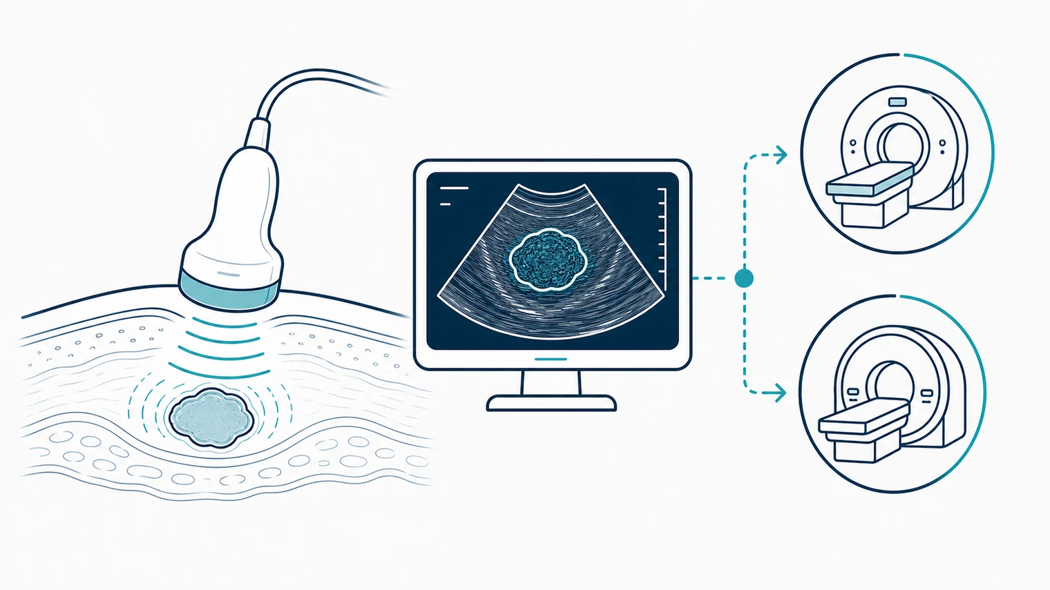
Where Ultrasound Stops
One thing worth knowing: no imaging test confirms cancer on its own. Ultrasound, CT and MRI can all flag suspicious masses and describe them in detail, but the actual diagnosis comes from a pathologist looking at tissue under a microscope.
When ultrasound flags something suspicious, the next steps usually include:
- Further imaging like MRI or CT
- A tissue biopsy for a firm diagnosis
- A specialist appointment to talk through what was found
What to Expect on the Day
A good sonographer will walk you through everything before they start, because anxiety makes the whole appointment harder than it needs to be.
Before the Scan
Preparation depends on what's being scanned. Some abdominal scans need fasting beforehand, pelvic scans need a full bladder, and others don't need any preparation at all. You'll get the instructions specific to your scan when you book in.
During the Scan
A water-based gel goes on the skin to remove the air between the probe and your body. The gel can feel cold, although we warm the bottle first where possible. You stay awake throughout, and the sonographer might ask you to hold your breath now and then to keep the image still. The probe needs firm pressure in some areas, which shouldn't hurt but you will feel it.
Most scans run between 20 and 30 minutes, with complex cases taking a little longer.
After the Scan
Results usually don't come on the same day, because the images need a specialist to review them properly. The wait can feel hard, but rushing the read is worse than the wait itself.
Worth repeating: ultrasound can spot a suspicious change, but only a biopsy confirms cancer. The faster something gets flagged, the faster you can get in front of a specialist and start a treatment plan if you need one.
Accuracy, False Positives, False Negatives
No scan is perfect, and it's worth being honest about that.
False Positives
A false positive happens when the scan flags something suspicious but there's no cancer there. Benign findings can look frightening on screen, which is stressful in the moment, although the follow-up testing usually clears things up within a week or two.
False Negatives
A false negative is the scarier scenario, where the scan looks clean but a tumour is actually present. That's why a lump you can feel shouldn't be dismissed simply because the ultrasound came back clear. If something is worrying you, push for further testing, because a clean scan is rarely the last word.
Ultrasound Earns Its Keep at Biopsy
When something suspicious does turn up, a biopsy is almost always needed to make the call. Ultrasound has another role to play here, because it's used in real time to guide the biopsy needle precisely to the target site. You stay awake, the needle is watched on screen as it goes in, and the whole procedure usually runs between 15 and 30 minutes.
Mobile Ultrasound in Brisbane
At Modia Health we bring the ultrasound to you, whether that's at home, in an aged-care facility, or somewhere else that suits. It means skipping the hospital trip, the parking, and the waiting room.
Our portable scanners run high-resolution imaging with Doppler, and our sonographers are trained across tumour assessment, lymph nodes and soft-tissue work. You'll often get a preliminary read on the day, and the full digital report goes to your GP within 24 hours so nothing gets lost in handover.
When to Get Checked, and When to Move Fast
Some signs need to be looked at the same day, while others can wait for a routine scan in the next week or two.
See a doctor today if you have:
- A lump that's growing quickly
- Hard lymph nodes that don't shift when you press them
- Severe abdominal pain you can't explain
- Weight loss without an obvious reason
Book a routine scan if you have:
- A new lump you can feel
- Lymph nodes that stay swollen for more than a couple of weeks
- A family history of cancer
- Persistent symptoms your GP wants a closer look at
Your Next Steps
If something is worrying you, the best thing you can do is not sit on it. Finding things early saves lives, and ultrasound is one of the fastest, safest ways to get a first look at what's going on.
Our mobile ultrasound team covers Greater Brisbane and the surrounding regions, bringing hospital-grade imaging to wherever works for you. Give us a call when you're ready, and we'll make the rest of it as straightforward as possible.
More from Modia Health:
Disclaimer: This article is general information and isn't a substitute for medical advice. Please talk to a qualified clinician about your own situation.